What makes up a true neural stem cell?
What controls its self-renewal and commitment?
How is the symmetry of division regulated and kept in check?
Can we pinpoint the origin of brain cancer?
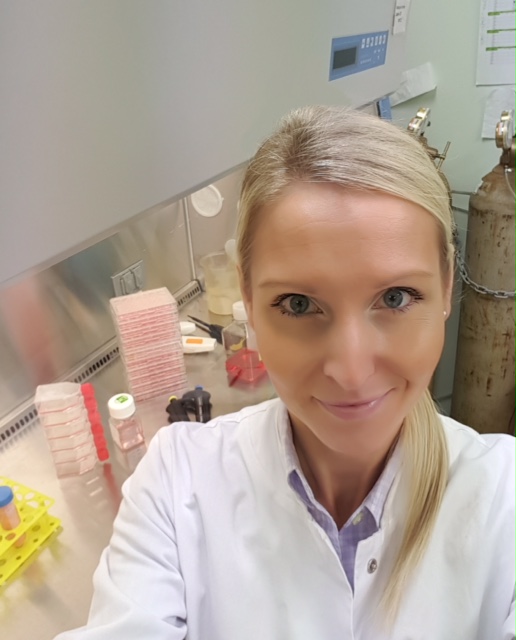
Questions about molecular mechanisms behind the regulation of cell fate within the mammalian brain have been fascinating scientists around the world for centuries. The effort to address the essential topics is not only a delightful and captivating everyday struggle of neuroscientists but it’s also a part of a global movement and a world-scale attempt to improve and fight for lives of people touched with neurodegenerative diseases, neurotrauma and brain cancer. In Porter Lab, we have that great privilege to contribute to this global effort. The Brain Group, as the vital organ of our lab, is committed to high-quality research, breakthrough discoveries and new, exciting ideas.
Several of The Brain Group projects are focused on the role of a cyclin-like protein Spy1, aka Speedy-1, SPDYA, RINGO A, in neurogenesis and neural types of cancer. We know that Spy1 activates CDK1 and CDK2 in a unique way and promotes the degradation of the CDK inhibitor, p27Kip1. Since CDK2 and p27Kip1, play a regulatory role in many developmental events including neurogenesis and these effectors are aberrantly regulated in several aggressive forms of cancer like glioma, we consider investigating Spy1 function of high importance.
NTA Joint Venture
Our brilliant Ph.D. candidates, Ingrid Qemo and Frank Stringer, are the main investigators of Spy1 role in neurogenesis and expansion of neural stem cells. Based on studies of other neural systems in the lab, like neuroblastoma, we’ve learned that Spy1’s cell cycle-mediated effects are key factors in regulating terminal differentiation in neurons. In addition, examination of the developmental time course of the mammalian brain revealed that significant decline of Spy1 levels coincides with events of neuronal differentiation and apoptosis, whereas the peak of expression overlaps with the phase of increased stem cell proliferation.
Now, a question arises about the importance of Spy1 in maintaining pools of adult neural stem cells and potential consequences of its aberrant regulation.
To address this question we were successfully funded by Canadian Cancer Society, initially with Innovation, and later on, with Innovation to Impact grants which allowed us to generate tools and purchase sophisticated equipment required for detailed and high-quality investigation.
Although one of our dedicated undergraduate students, Dalton Liwanpo, is yet to pinpoint specific neural cell populations abundant in Spy1 protein and mRNA, Ingrid and Frank are already utilizing an inducible transgenic NTA mouse model (Nestin-Spy1-pTRE mouse) to obtain upregulation of Spy1 protein in select, Nestin+, populations of neural stem cells. So far, this system successfully allowed for investigation of the spatiotemporal function of Spy1 in vivo. Ingrid’s expertise in primary cell cultures and Frank’s proficiency in immunoassays on tissue sections, which is currently being passed on to Dalton, came together to establish ample data containing surprising results and exciting phenomena, currently put together in a manuscript to be shortly published as an original article.
Although to date, we have not observed brain tumours in those mice, Spy1, due to its unique functions, constitutes a perfect accessory to ”the crime of brain tumourigenesis”, and this is the main focus of currently ongoing studies and in part a leading objective of another undergraduate project thesis by a diligent student and an Outstanding Scholar, Youshaa El-Abed.
The Glioma Enterprise
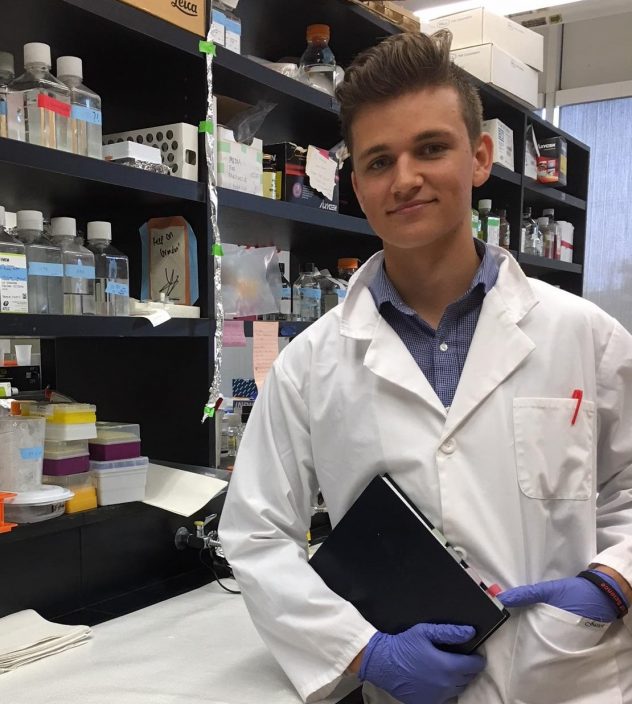
In spite of the fact we are yet to discover where the story of Spy1 in brain tumourigenesis starts and ends; whether Spy1 is a factor during initiation or a promising therapeutical target, we did establish its role in glioma maintenance and progression. The funding obtained from Cancer Research Society was crucial in the initial groundbreaking phase of this study. With an amazing support of the co-authors and our collaborators, I was finally able to share six years worth of data in a high impact study, Cancer Cell 25:64-76. We utilized primary cell cultures derived from GBM (Glioblastoma Multiforme) patients and isolated CD133+ cell populations, known to have an extremely high capacity of glioma formation upon orthotopic injection into mice. The depletion of Spy1 levels in those cells caused a significant increase in differentiation markers and enhanced frequency of tumorsphere formation. Further investigation revealed a striking effect of Spy1 depletion on the mode of division of CD133+ cells; we found that decline in Spy1 levels caused a highly significant increase in the rate of asymmetric divisions. We are currently building upon this exciting data, trying to further dissect the heterogeneous cell populations of GBM in respect to its established subtypes. Alex Rodzinka, who is an outstanding and a very bright undergraduate student, awarded with the Brain Tumour Foundation of Canada Research Studentship, is attempting to shed light on the essentiality of Spy1 in the expansion of those populations and targeting Spy1 for their potential eradication, which is tested using Zebrafish platform.
Another burning aspect, awaiting answers, is the mechanism behind our results; although we established WHAT it is, that Spy1 is doing in those GBM cells we still don’t know HOW. Mat Stover is The Brain Group’s only MSc student, his persistence and dedication are exactly what’s needed to take on this task. In addition to molecular pathway determination, Mat is studying how changes in Spy1 levels affect cell cycle profile in glioma subpopulations and how we can utilize that modulation for therapeutic purposes.
The complexity of GBM makes it extremely hard to treat and ironically that complexity expands as our knowledge about it advances, hence, the therapeutic progress always ends up behind. Can we win this race? Current literature suggests that the patient-tailored treatment has the potential for best clinical outcomes. Now, to determine individual approach, should the patients be evaluated genomically only? Or proteomically, phosphoproteomically, metabolomically or by the cell subpopulation composition? Should we search for additional tools to predict the response of a tumour to therapy?
Tumour Stress Alliance
Our collaborators from Henry Ford Hospital (USA) have developed imaging method (DCE-MRI) module to measure brain tumour physiology and tissue properties such as cellularity, Extracellular space, and perfusion.
Patients presenting with low rates of tumour perfusion due to solid stress (pressure from solid components of a tumour) causing extensive hypoxia and impaired drug delivery, exhibit poorer prognosis with worse chemotherapy response and shorter survival in comparison to patients with high tumour perfusion. Therefore, a tool allowing for measurement of the tumour solid stress and for subsequent therapy response prediction could make a substantial difference.
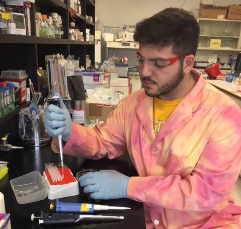
In our lab, we were able to develop, optimize and validate a 3D platform mimicking patient brain tumour growth in vitro. This system offers an extensive flexibility of environment manipulation and a high throughput approach for drug testing, which are impossible in vivo. Therefore this model has become an in vitro approach to study and compare, to the results obtained through DCE-MRI, the biology of the tumour under solid stress. Jonathan O’Beid is a very enthusiastic undergraduate thesis student who has gained ample of experience on immunostaining of DCE-MRI- assessed brain tumour tissues and analysis of the expression of crucial stress related marker proteins. Jonathan is utilizing the 3D system to manipulate solid stress-related factors and study their effect on drug response in vitro.
Due to the fact that our 3D system allows to entirely control tumour growth and to establish a high throughput time course, we are able to collect data on changes to the cellular layers of our tumour model. The results can be utilized to mathematically model tumour growth before, during and post-therapy and how the arising internal solid stress affects the cellular composition. We are currently in process of collecting and analyzing data which will be forwarded to our collaborators at the Department of Engineering. They are motivated to establish a mathematical approach to the brain tumour growth and drug response prediction.
Our novel 3D platform is fully patient customized and, in combination with the above unconventional interdisciplinary methods, it can, not only, potentially move forward our knowledge on GBM biology but also become a drug response prediction tool, pointing at the necessity of diverse fields of science to come together in understanding to tackle the most complex phenomena.
Medulloblastoma Incorporated
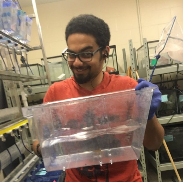
The medulloblastoma story is slowly coming together as it is one of the projects in our lab which are being passed from hands to hands of very keen and skilled undergraduate students who take over, one after another, bringing all the pieces together at the end. This project was funded by Brain Tumour Foundation of Canada which also generously offered several Research Studentships related to medulloblastoma studies in our lab.
Over the years we have discovered that medulloblastoma cells cultured as neurospheres enriched in Spy1 but not in Cyclin E1, suggesting a unique role of Spy1 in this particular tumour. Utilizing in vivo zebrafish assays we found that Spy1 levels downregulation is essential in increasing efficacy of medulloblastoma response to synthetic CDK inhibitors.
Philip Habashy, who took over this project, performed further analysis of FACS derived stem-like medulloblastoma cell populations and revealed that Spy1 is an important factor in maintaining their character. Interestingly, Phil’s curiosity made him shift gears recently and work on a novel idea, together with Dr. Huiming Zhang (University of Windsor, Biology), trying to address the role of GABAB receptor activity in the proliferation of medulloblastoma cells.
The Brain Group’s mission is to dissect mechanisms responsible for control over the fate of neural stem cells and progenitors utilizing diverse in vitro and in vivo systems. The data obtained on Spy1 role in glioma suggest that this unique protein has a potential to become an important therapeutic target. Overall we hope to contribute to better understanding of brain cancer biology and finding new potential ways to target tumours in a specific and efficient way.
I hope you enjoyed getting to know a bit about the Porter’s Lab Brain Group; let us know your thoughts in the comments below!
Dorota Lubanska, Ph.D

Pingback: Porter Lab Year in Review | Porter Lab