Our amazing
Facilities.
We are well equipped.
Switch the toggle
- User fee – $ 25.00/hour – annual charge.
- Cap fee – $ 1000.00 for 40 to 100 hrs and $ 2000.00 for more than 100 hrs per year.
- Overnight timelapse imaging – $ 75.00 for an experiment.
- Training fee – $ 25.00 per person – Training session is done during the first week of each month.
- The room CORe-104 is BSL2. Please, check the SOP binder located in the room before using the scope.
- No radiolabeled material is permitted.
- No food or drink is permitted inside the room.
- No gloves should be worn when operating the scopes.
- For more information and for booking training please contact Dr. Elizabeth Fidalgo.
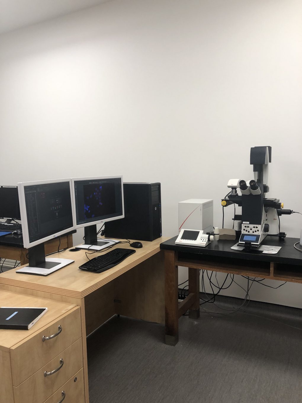
Leica scope
The Leica DMI6000 fluorescent microscope can be used for a variety of applications in fluorescence microscopy and image analysis, including time lapse experiments. It has YFP, CFP, GFP, Texas Red, Cy5, A4 filter cubes. It is supplied with the LAS and LAS AF soltware. Objectives include: Dry (2.5x, 10x, 20x and 40x) and Oil (40x and 60x/100x). The Leica DMI6000 has a colour camera in addition to the fluorescence camera and can be used for bright field, DIC, and phase contrast as well. Individuals who wish to use the Leica scope must contact Dr. Elizabeth Fidalgo for more information and training.
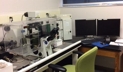
Confocal scope
e confocal microscope, a Biorad 1024 on a Nikon Eclipse 800 microscope, has the capability of combining double label imaging with DIC transmission optics. It has a krypton/argon laser with excitation at 488 nm, 568 nm and 647 nm. Objectives include 20x, 40x oil and 60x oil. The software includes quantitative analysis for distance,staining intensity and double-label co-localization. Triple label sequential analysis is also available. A cryostat (Micron) is available for preparing sections for confocal imaging. Individuals who require confocal image analysis in their research advised to contact Dr. Andrew Swan for more information
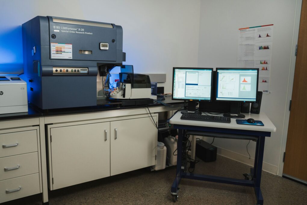
BD LSR Fortessa™ X‐20
- 488 nm (Blue)
- 640 nm (Red)
- 405 nm (Violet)
- 561 nm (Yellow/Green)
- 355 nm (UV)
- Blue ‐ FITC (530/30BP, 505LP), PerCP Cy5.5 (695/40BP, 685LP)
- Red ‐ APC (660/20BP), AF700 (730/45BP, 695LP), APC‐Cy7/APC‐H7 (780/60BP, 750LP)
- YG – PE/DsRed (582/12BP), PE‐TR/mCherry (610/20BP, 550LP), PE‐Cy5 (670/30BP, 635LP), PE‐Cy5.5 (710/50BP, 695LP), PE‐Cy7 (780/60BP, 750LP)
- Violet ‐ BV421/Pac Blue (450/50BP), BV510/AmCyan (528/50BP, 475LP), BV605 (610/20, 595LP), BV650 (670/30BP, 635LP), BV711 (710/50BP, 695LP), BV786 (780/60BP,750LP)
- UV – Indo‐1 (violet)/AF350/DAPI/Hoechst (450/50BP), Hoechst Red/Side Population (675LP)
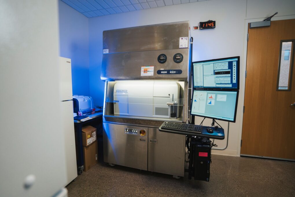
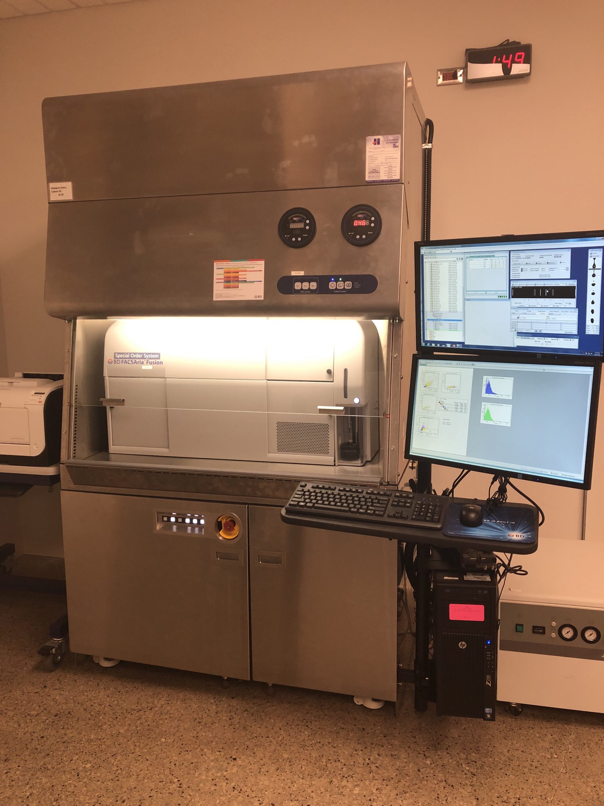
BD FACSAria Fusion™
- 488 nm (Blue)
- 640 nm (Red)
- 405 nm (Violet)
- 561 nm (Yellow/Green)
- 355 nm (UV)
- Blue – FITC (530/30BP 502LP), PerCP‐Cy5.5 (695/40BP 655LP)
- Red – APC (660/20BP), APC‐Cy7 (780/60BP 735LP), AF 700(760/45BP, 690LP)
- Violet – BV421/Pac Blue (450/40), BV510/AmCyan (510/50BP, 502LP), BV 605 (610/20BP, 600LP), BV650 (660/20BP, 630LP), BV650 (670/30BP, 635LP), BV711 (710/50BP,
695LP) - PE/DsRed (582/15BP), PE‐TR/mCherry, PI (610/20BP 600LP), PE‐Cy5 (670/30BP, 635LP), PE‐Cy5.5 (710/50BP 630LP), PE‐Cy7 (780/60BP 735LP)
- UV – Indo‐1 (violet)/AF350/DAPI/Hoechst (450/50BP), BUV395 (378/28BP)
FlOW CYTOMETRY INFO
- All requests for analyzing and sorting samples must be accompanied by the Biosafety
form, sign by the principal investigator (PI) - BSL2 samples must be fixed before they are analyzed on the cytometer, these samplesinclude: All infectious, human, and non-human primate cells, cells manipulated with viralagents (EBV, vaccinia, HTLV, lentivirus, nanoparticle, etc). If the in vivo model is
considered BSL2 then your sample is a BSL2 sample.
- Primary human samples used for sorting need to have the screening results and benegative for: Hepatitis B, C, HIV, Mycobacterium tuberculosis, Neisseria meningitides,Chlamydia psittacci, Coxiella burnetii, HTLV-1,2, LVMV, vesicular stomatitis virus
infections.
- Samples manipulated with viral agents: EBV, vaccinia, HTLV, lentivirus, nanoparticle,etc must be clearly described in the Biosafety form. These samples need to be washed atleast 3 times and passed through a 70 micron nylon filter just prior to being brought to this
facility.
- Gloves should be worn by all users at all times.
- No food or drink is permitted.
- During sign-up procedure users must notify the flow facility manager about the biosafetylevel of the material to be analyzed or sorted.
- No radiolabeled samples are permitted on any instrument.
- All biological waste must be removed from the facility for disposal in their homelaboratory.
Ethanol-fixation of samples
- Place 1 x 10^6 cells into a polypropylene tube and centrifuge at 1000 rpm for 5 min.
- Resuspend the pellet with 1 mL of PBS (1x) and centrifuge at 1000 rpm for 5 min
- Remove the supernatant without disturbing the pellet and add (dropwise) 1 mL of -20 C 70% EtOH to the cell pellet while vortexing.
- Keep cells on ice for 2 hrs or at -20 C until the day of the staining (cells can be stored for several weeks at -20 C)
- On the day of staining, centrifuge the samples at 1000 rpm for 5 min, remove the supernatant
- Resuspend the pellet in 1 mL of PBS (1x), centrifuge again, remove the supernatant
- Resuspend the pellet in 0.5 mL of PBS (1x) + 2 mM EDTA and proceed to the staining protocol
- DNA staining with PI (The facility doesn’t run samples with PI anymore, please check the DAPI staining protocol below)
Cell viability with DAPI – BD website
Staining of Live Cells for Viability Analysis by Flow Cytometry
1. Obtain a single-cell suspension.
2. Resuspend cells in BD Pharmingen™ Stain Buffer (FBS) (Cat. No. 554656) or 1× Dulbecco’s Phosphate Buffered Saline (DPBS) containing 0.05-0.2 μg/mL DAPI.
a. The optimal concentration of DAPI for viability analysis may vary by cell type. We recommend titrating the reagent for your cell type of interest in early experiments.
b. Additionally, apoptotic cells may stain with variable amounts of DAPI. If further analysis of apoptotic cells is desired, we recommend co-staining with BD Pharmingen™ FITC Annexin V (Cat. No. 556419).
3. Incubate for 5 minutes at room temperature. No wash is necessary before analysis.
4. Proceed to analysis by flow cytometry.
DNA staining with DAPI – BD website
Staining of Fixed Cells for DNA Content Analysis by Flow Cytometry
1. Obtain a single-cell suspension.
2. Treat cells on ice for 30 minutes with 70-80% ice-cold ethanol.
a. Ethanol fixation typically provides the most resolved histograms. However, this reagent has also been successfully used for DNA content analysis with the Transcription Factor Buffer Set (Cat. No. 562574/ 562725) or BD Cytofix™ Fixation Buffer (Cat. No. 554655) and BD Phosflow™ Perm Buffer III (Cat. No. 558050) protocol.
3. Wash cells once with BD Pharmingen™ Stain Buffer (FBS).
4. Dilute DAPI solution to 0.5-1 μg/mL in Stain Buffer (FBS) or 1× DPBS immediately before use.
5. Stain cells for 5-15 minutes at a 1 – 2 x 10^6 cells/mL cell density. No further wash is necessary before analysis.
a. The optimal cell density and concentration of DAPI for DNA content analysis may vary by cell type. For best results, assay conditions should be optimized in early experiments.
6. Proceed to analysis by flow cytometry.
Buffers for cell sorting and analysis
1x PBS (Ca2+/Mg2+ free)
0.5 – 2 mM EDTA
1-2% FCS (only for sorting)
0.2 µm filtered, stored at 4 °C
Or if pH-stability is crucial
1x PBS (Ca2+/Mg2+ free)
1 mM EDTA
25 mM HEPES pH 7.0
1% FCS (heat-inactivated) or BSA
0.2 µm filtered, stored at 4 °C
Cell surface antibody staining – BD Stain Buffer-Source: cat# 554656
Intracellular antibody staining – BD Pharmingen: cat# 562574
Add 1 uL of DAPI solution 0.5-1 μg/mL) to 0.5 ml of PBS (1x) + 2 mM EDTA containing 1 x 10^6 cells or less, and keep the samples for at least 20 min on ice and covered. Use a 5 ml Falcon BD Flow tube.
Facility manager – Dr. Elizabeth Fidalgo da Silva
Facility hours of operation – Analyzer – Tuesday, Wednesday and Thursday – 9:00 am – 5:00 pm
Sorter – Tuesday and Wednesday – 9:00 am – 5:00 pm
*9:00 – 10:00 daily – Instrument setup and 4:00 – 5:00 – Instrument clean up
*Booking window – Up to one week in advance
*Off-hours facility access – PIs only
*Facility fees are used for necessary maintenance, software updates, setup and flow supplies
- All fees are per hour
- Training is individual
- Experiments run by the facility. The facility manager will aid in the experimental setup. The facility manager will run the sample and provide complete results.
- Assisted experiments – The facility manager will be by your side throughout the experiment and will assist with the setup. The facility manager will perform cleaning and shut down of the instrument. *Trained individuals or individuals who have not run an experiment in 3 months require assisted runs before using the machine unassisted (the number required is at the discretion of the facility manager, typically 1-2).
- Unassisted experiments – Trained individuals can run samples alone. Individual users are responsible for cleaning and shutting down the instrument.
| SORTER | ANALYZER | |
|---|---|---|
| OPERATOR | 120.00 | 100.00 |
| ASSISTED | N/A | 100.00 |
| UNASSISTED | N/A | 60.00 |
| CORPORATE | 200.00 | 160.00 |
| TEACHING | N/A | 100.00 (UNASSISTED) 120.00 (ASSISTED) |
| TRAINING | N/A | 100/person |
| DATA ANALYSIS | 80.00 | 100.00 |
| AFTER HRS | N/A | N/A |
our lab space
FOLLOW PORTER LAB ON TWITTER
@L_Porterlabrats
Copyright © 2021 Porter Lab