Our PASSION,
Our Research.
Areas of research interest.
Our tools
Our tools
our research programs and projects
Tumour Suppressor Program
Cell Cycle Regulation by Tuberin
The ability of cells to sense diverse environmental signals, including nutrient availability and conditions of stress, is critical for both prokaryotes and eukaryotes to mount an appropriate physiological response. Over the past three decades the proteins Tuberin, Hamartin and TBC1D7 have emerged as a large protein complex called the Tuberous Sclerosis Complex. This complex can integrate a wide variety of environmental signals to control a host of cell biology events including protein synthesis, cell cycle, protein transport, cell adhesion, autophagy, and cell growth. Porter Lab is interested in identifying the roles of Tuberin in the cell cycle regulation, specifically at the G2/M transition. For this purpose, we have created fluorescent tools that can be used in immunofluorescence and flow cytometry techniques. Click here to know more about our fluorescent tools.


Breast cancer program
Cell Checkpoint Regulation by Spy1
One of the major foci of our group is to elucidate the function and regulation of the human cell cycle protein Speedy (Spy1; SPDYA). Spy1 is an unusual cell cycle protein in that it can activate Cyclin Dependent Kinases (CDKs) in the absence of the typical post-translational modifications on the CDK. Spy1 binds directly to the G1/S CDK, CDK2, and the G2/M CDK, CDK1, to activate them and promote cell proliferation and oocyte maturation. Of major interest to us, this atypical activation of the CDK allows Spy1 to promote cell growth and division in the presence of quiescent/senescent stimuli.
Our lab has determined that Spy1 is elevated in several forms of human cancer including breast, brain, liver. While there are similarities in how we may target this protein in each of these systems, how it is functioning to drive cancer in each system is different and requires special models to study properly. Our breast program looks at how the Spy1 protein regulates normal breast development and what can go wrong with its regulation to result in susceptibility to cancers forming and progressing.
Brain cancer program
Brain cancer program is focused on the role of the cell cycle in the regulation of neurogenesis and neural types of cancer. We are interested in the control of the mode of division in normal neural stem cells and how disruption of the symmetry/asymmetry balance at the cell cycle level contributes to aberrant growth at initiation and during progression of glioblastoma (GBM) the most aggressive type of brain cancer. Using high throughput design based on CRISPR-Cas9 technology we are able to perform in vivo screens in mice to elucidate mutations cooperating with cell cycle dynamics and signatures driving specific mode of division profiles.
Several projects of the Brain cancer program are dissecting the intratumoural dynamics between individual populations of Brain Tumour Initiating Cells (BTICs) and the cell population- specific mechanisms developed to resist current therapeutic approaches leading to tumour recurrence and relapse in patients with GBM. Patient- disease customized 3D Brain Tumour Organoid model, developed in our lab, in conjunction with NGS and live cell studies, allows us to investigate biology of GBM in the context of its microenvironment and how its components regulate BTIC maintenance and drug resistance in real time. To move therapeutic efforts forward, we intensively collaborate with the Chemistry Department to seek new treatment strategies against glioblastoma and novel, breakthrough compounds and systems are being evaluated using both the organoid as well as Zebrafish PDX platform. Over the years, Brain program has developed multiple valuable local and international collaborations which allowed us to access GBM patient tumour specimens. With, fully evaluated at the genomic and functional level, live material obtained at the surgery we started a GBM biobanking initiative in our lab which has been supporting the life relevance of our brain related studies.


Liver cancer program
Hepatocellular carcinoma (HCC) is one of the leading causes of cancer-related deaths worldwide, with a 5-year survival rate of only 20%. Many factors can contribute to the development of HCC such as viral infection, alcoholism, metabolic disorders and obesity. Advanced stages of HCC have no effective treatments available. A serendipitous discovery in our lab has revealed that the protein Spy1 (Speedy/Ringo; gene SPDYA) is capable of enhancing proliferation of liver cells and promotes the development of HCC. This research program uses mouse models as well has human cells to determine how this protein changes the cell biology that leads to the formation of cancer in the liver. Results from this work could reveal new avenues of treatment for this aggressive form of disease.
Prostate Cancer program
Prostate cancer (PC) is the second most common cancer in men worldwide, affecting 1 in 8 men worldwide. As treatment options continue to evolve, disease management and overall patient outcomes have improved. While effective, these treatments can sometimes pressure PC cells to transdifferentiate into a more aggressive, treatment-resistant type of PC, known as neuroendocrine prostate cancer (NEPC). Treatment options for NEPC are limited as it is resistant to all current therapies, leading to a poor overall prognosis with an estimated survival of less than one year. Our lab has identified a class of cell cycle regulatory proteins elevated in NEPC, with evidence supporting that these proteins have the potential to drive progression to this drug-resistant form of disease. Our work focuses on testing drugs that can inhibit these proteins and test whether these drugs can treat and/or prevent the progression of disease to NEPC. Preventing the progression of disease to NEPC and identifying markers of NEPC remains one of the greatest challenges in this field, and we have strong rationale and data to support this being a promising direction that could make a meaningful impact for PC patients.

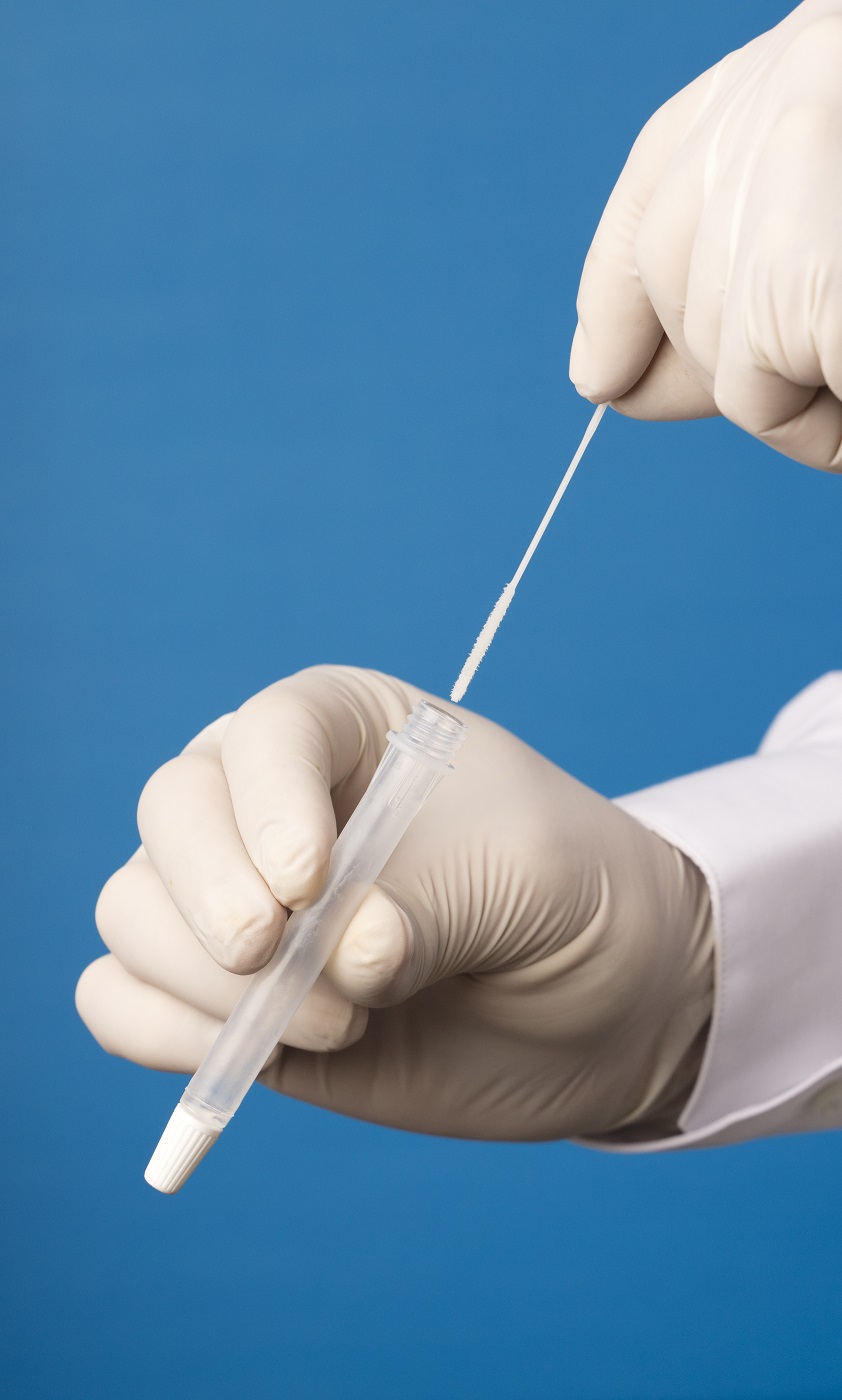
Covid screening program
This study involves a large multidisciplinary team focusing on: 1) detecting emerging SARS-CoV-2 variants, 2) providing molecular structural information to prioritize VOCs, 3) providing community-based surveillance to prevent COVID-19 outbreaks.
This work brings together three platforms: 1) Wastewater testing led by Dr. McKay. 2) Analysis of the structure of the SARS-CoV-2 proteins and the sequencing of variants led by Drs. Ng and Tong. 3) A saliva-based screening project being run by Drs. Porter, Tong, and Soucie.
Our diverse and multidisciplinary team uses our collective experience and expertise to move science and technology forward to rapidly detect COVID-19 outbreaks in a strategic Canadian land entry point. This work has the potential to provide invaluable information to regional and national public health networks and to support an efficient and economic model for rapidly detecting the emergence of novel viral species in our region. Long-term surveillance of this nature is the kind of forward thinking that we need to protect our Country against the devastation that we have faced with COVID over the past 2 years.
For more information, or to join the study, visit the web site: https://www.wesparkhealth.com/covid-screening-platform
a huge thank you
to our funders and supporters


Copyright © 2021 Porter Lab